
Three-dimensional (3D) spheroid assay is a crucial and powerful method to study the biological effects of cell model including cell proliferation, cell invasion and cell migration. This technique based on the 3D structure of spheroid has more physiological tissue-like morphology and close cell-cell contacts which could mimic cell interaction In Vivo. This distinct characteristic of three-dimensional spheroid assay has many advantages in researching the life activities of organisms. In addition, this assay can mimic different statuses of nutrient and oxygen supply. For instance, the migration of tumor cells out of small cancer cluster can be closely mimicked and cell migration of tumor cells researched by this method is useful for us to understand the real situation In Vivo. The underlying mechanisms of different types of cell migration or cell invasion In Vivo also can be further researching.
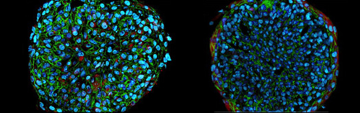 Figure 1. The diagram of spheroids coated with cells
Three-dimensional (3D) spheroid assay provide a rapid and automate cell migration and cell invasion system in a highly reproducible, standardized methods. Multicellular spheroids are coated with cells and placed on the top of a conventional cell culture dish or in a 96 well-plate form as one spheroid per well. After the spheroids incubated with culture medium for a period of time and attachment of the spheroids to the plastic surface, the cells that have migrated from the spheroids can be monitored and cell movement can be measured by living cell microscope. This technique is usually used to study the influences of some substances to cell migration including pro- or anti-migration such as the inhibition effects of tissue factor pathway inhibitor-2 to cell migration and cell invasion in prostate cancer cells.
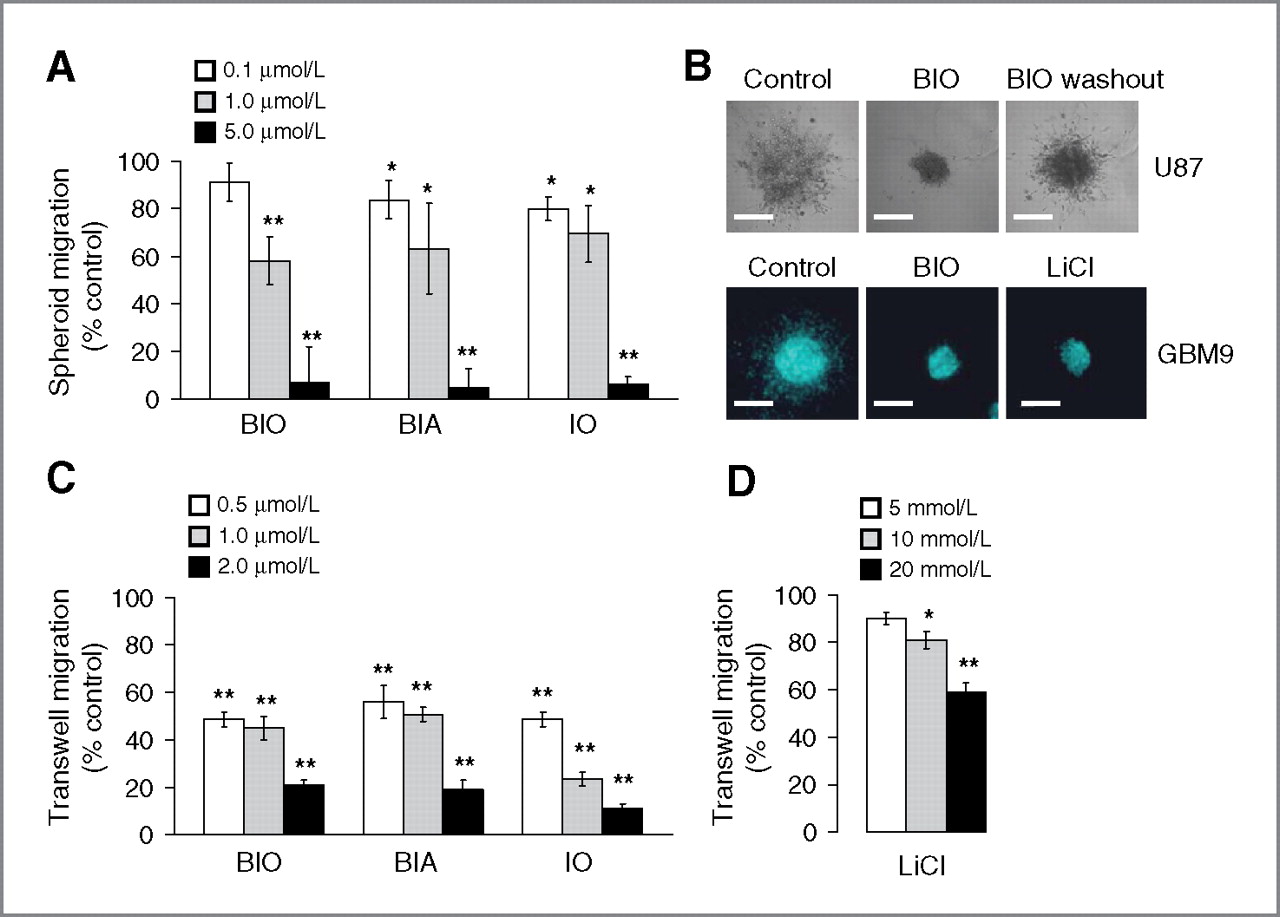 Figure 2. The experimental results of three-dimensional (3D) spheroid assay
Spheroid assay at Creative Bioarray not only offers all advantages mentioned above, but also allows for research on cell-cell interaction. Cells coated with spheroids are labeled with fluorescence such as cells are modified to express fluorescent proteins like GFP protein. One type of cells is cultured in plastic dish or 96-well plate, the other type of cells is coated with spheroids. Upon attachment, cells coated with spheroids can interact with the monolayer cells. The close interaction ensures paracrine crosstalk and cell-cell interaction. This platform allows us to analyze the migration changes influenced by the interaction between the two types of cells. Cells labeled by fluorescence can be easily monitored, which ensures the accurate data analysis of cell migration.

*If your organization requires signing of a confidentiality agreement, please contact us by email.
Online Inquiry