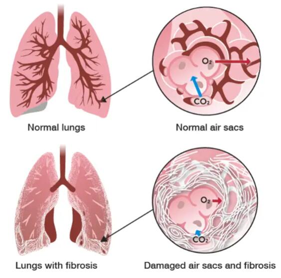
Lung fibrosis, also known as pulmonary fibrosis, is a chronic lung disease characterized by the formation of excessive scar tissue in the lungs. This can lead to breathing difficulties and a decreased ability to exchange oxygen and carbon dioxide. It can be caused by various factors, such as exposure to certain toxins, infections, or autoimmune diseases.
 Figure 1. Reprinted from Nature Reviews Disease Primers, 3, Martinez FJ, Collard HR, Pardo A, et al. idiopathic pulmonary fibrosis, 17074, 2017, with permission from Springer Nature.
In drug screening for lung fibrosis, in vitro cell models can be used to evaluate the efficacy and safety of potential therapeutic drugs. Creative Bioarray offers a TGF-β-induced fibrosis model using lung cells for researchers studying pulmonary fibrosis and related diseases. Researchers can then expose these cells to various compounds, including potential drug candidates, to assess their effect on fibrosis-related processes.
 Fibroblast-to-myofibroblast transition (FMT)
 Epithelial-to-mesenchymal transition (EMT)
Study Examples:
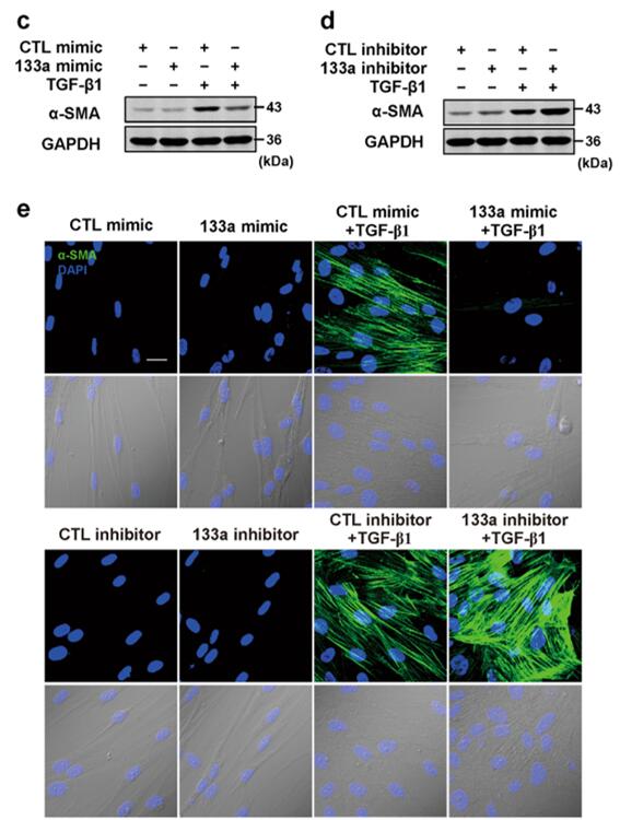
 Figure 2. TGF-β1-induced miR-133a blocks pulmonary fibroblast differentiation. [2]
References:
1. Martinez, Fernando J et al. "Idiopathic pulmonary fibrosis." Nature reviews. Disease primers vol. 3 17074. 20 Oct. 2017, doi:10.1038/nrdp.2017.74
2. Wei, Peng et al. "Transforming growth factor (TGF)-β1-induced miR-133a inhibits myofibroblast differentiation and pulmonary fibrosis." Cell death & disease vol. 10,9 670. 11 Sep. 2019, doi:10.1038/s41419-019-1873-x
Online Inquiry