
In the study of autophagy, electron microscopy is indispensable. The results obtained by other methods often need to be verified by electron microscopy. Some specific types of autophagy and ultrastructure of organelles also need to be observed by an electron microscope. Creative Bioarray has a professional instrument platform and experienced researchers who have been engaged in cell biology research for many years.
Autophagy is a lysosomal-dependent degradation pathway widely present in eukaryotic cells. Intracellular damaged proteins and organelles are degraded through autophagy, and the degradation products such as amino acids and fatty acids are then reused by cells. Stresses such as growth factor deficiency, oxygen-free radicals, and DNA damage can all induce autophagy. Autophagy plays an important role in cell growth and development, cell death, and some disease processes, such as tumor occurrence, development and treatment response.
Creative Bioarray has been committed to cell biology technology research for many years. Based on advanced technical equipment and complete platform construction, we can provide customers with a variety of reliable autophagy detection services. Our services include but are not limited to:
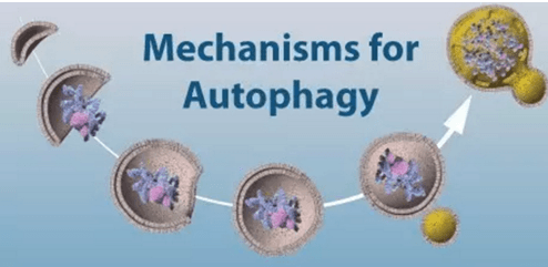
We use an electron microscope to observe the samples and determine whether there is an autophagic bubble structure. Under the electron microscope, damaged organelles such as swollen and degenerated mitochondria can be observed in autophagy cells. There is a bubble-like double membrane-like structure around it, or a double membrane surrounds the mitochondria to form autophagosomes. Residues that cannot be degraded in the lysosome can also be observed in autophagy cells.
This method can characterize autophagy based on the presence or absence of autophagic vesicles in the sample. Researchers need to have a clear understanding of the structure of autophagic vesicles, so as not to confuse autophagic vesicles with other types of vacuoles. Creative Bioarray has a professional instrument platform and experienced researchers who have been engaged in cell biology research for many years to provide you the most reliable service on autophagy detection.
In short, electron microscopy is a direct method for detecting autophagosomes, and it is also the gold standard for detecting autophagosomes, especially suitable for the first discovery of autophagy induced by a certain stress condition.
Service Process

Applications
Autophagy plays a very important role in the body's immunity, infection, inflammation, tumor, cardiovascular disease, and neurodegenerative diseases. At present, the three most prevalent diseases in autophagy research are tumors, neurodegenerative diseases and immune diseases.
The time depends on the experiment content
If you are interested in our services, please contact us for more detailed information.
Online Inquiry