Resistance to apoptosis, a natural cell death process, is a significant factor in chemotherapy failure during cancer treatment. However, finding a way to bypass apoptosis and induce cancer cell death through alternative pathways shows promise in overcoming this issue. Necroptosis offers an alternative mechanism for programmed cell death, effectively addressing apoptosis resistance and potentially enhancing the immune response against tumors in cancer therapy.
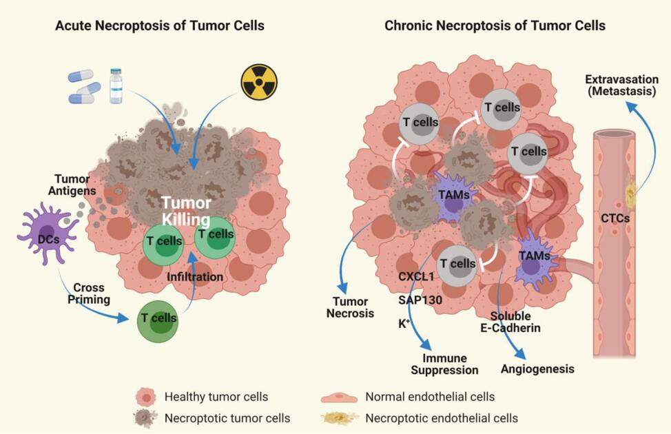 Figure 1. Role of Necroptosis in Cancer. [1]
Necroptosis is initiated through the ligation of TNF receptor family proteins (TNFR, FAS, TRAILR, and DR6) with RIPK1–RIPK3 when Caspase-8 activity is impeded. Additionally, activation of TLR3 and TLR4 by double-stranded RNA (dsRNA) and LPS in macrophages, respectively, can trigger necroptosis through TRIF-dependent activation of RIPK3. Viral RNA and damaged mitochondria can trigger necroptosis by activating RIPK3 through ZBP1. Once activated, RIPK3 phosphorylates MLKL, causing it to form oligomers. These oligomerized MLKL molecules then move to the plasma membrane, where they interact with ion channels and induce rupture of the membrane.
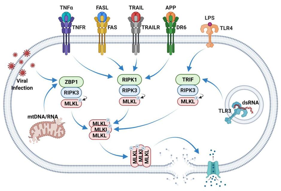 Figure 2. Overview of Necroptosis Pathway. [1]
Creative Bioarray offers a cutting-edge necroptosis assay, providing a comprehensive solution for researchers studying necroptosis in cancer cells.
Detecting Hallmarks of Necroptosis
- Loss of Membrane Integrity
LDH Release Assay: The lactate dehydrogenase (LDH) release assay is commonly used to measure the release of LDH into the culture medium, indicating loss of membrane integrity and necrotic cell death.
- Morphological Changes
Light Microscopy and Electron Microscopy: Necroptotic cells often display characteristic morphological changes such as cell swelling, plasma membrane rupture, and a "necrotic" appearance. These changes can be visualized using light microscopy and confirmed at the ultrastructural level with electron microscopy.
- Key Protein Detection
Immunoblotting: Detection of key necroptosis-associated proteins, including receptor-interacting protein kinase 1 (RIPK1), RIPK3, and mixed lineage kinase domain-like protein (MLKL), can be performed through immunoblotting. Phosphorylation of MLKL is a crucial event in necroptosis.
Immunofluorescence Staining: Visualization of necroptosis markers within cells using immunofluorescence staining provides spatial information about the distribution of key proteins, confirming the activation of necroptotic pathways.
- Cell Viability and Cytotoxicity Assays
MTT Assay: The MTT assay measures cellular metabolic activity and can be used to assess changes in cell viability associated with necroptosis.
Propidium Iodide (PI) Staining: PI is a membrane impermeable dye that stains necrotic cells with compromised membrane integrity. Flow cytometry analysis can quantify PI-positive cells.
- Death Receptor Activation
TNF Treatment: Necroptosis is often triggered by the activation of death receptors, particularly tumor necrosis factor (TNF) receptors. Treatment with TNF can induce necroptosis, and its effects can be blocked using specific inhibitors.
- Genetic Approaches
siRNA or shRNA Knockdown: Targeted knockdown of necroptosis-associated genes using small interfering RNA (siRNA) or short hairpin RNA (shRNA) can help confirm the role of specific genes in necroptosis.
References:
1. Yan J, Wan P, Choksi S, Liu ZG. Necroptosis and tumor progression. Trends Cancer. 2022;8(1):21-27. doi:10.1016/j.trecan.2021.09.003
2. Vanden Berghe T, Grootjans S, Goossens V, et al. Determination of apoptotic and necrotic cell death in vitro and in vivo. Methods. 2013;61(2):117-129. doi:10.1016/j.ymeth.2013.02.011
* For scientific research only
