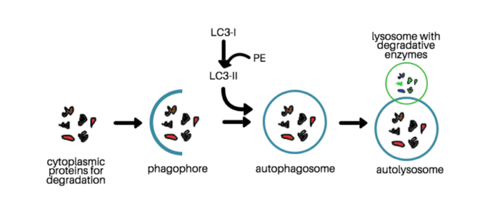
Autophagy is related to cell growth, development, death and many human diseases (such as cancer, neurodegenerative diseases, liver diseases, heart disease, infections and immune diseases). Therefore, autophagy is very important in life sciences. Creative Bioarray provides several dynamic detection services to study autophagy.
In recent years, methods for detecting autophagy continue to emerge.However, no method can detect autophagy with absolute accuracy and specificity.Therefore, it is recommended to combine multiple methods to obtain reliable data.Combining dynamic and static detection methods can more effectively explain the induction of autophagy and changes in autophagy flux.
Autophagy flux detection is a detection method that treats the entire autophagy process as an object. Autophagic flux refers to the complete process of autophagy, including the transport of the package formed by the autophagosome to the lysosome, and the fusion with the lysosome to form an autophagic lysosome, as well as the degradation and recycling of the package.
The detection method of autophagic flux is more complicated, and it is often impossible to systematically detect autophagic flux using one of the existing technical methods alone. Therefore, it is necessary to choose a variety of different experimental methods and comprehensively evaluate the detection results to reflect the state of autophagy flow more objectively.

Dynamic detection can prove the existence of autophagy and reflect the entire dynamic process by detecting the degradation rate of autophagic proteins or the accumulation of autophagosomes. Our services include but are not limited to:
Use Autophagy-related Drugs to Detect LC3B-I/II Conversion Service
Autophagy is a highly dynamic, multiple-step process that can be modulated at several steps. The amount of autophagosomes detected at any specific time point is a function of the balance between the rate of their generation and the rate of degradation through fusion with lysosomes. Accordingly, increased LC3-II could reflect either increased autophagosome formation due to autophagy induction, or a blockage in the downstream steps in autophagy. The mere detection of levels of LC3-II at a specific time point is insufficient for an overall estimation of autophagic flux, which refers to the complete process of autophagy including the sequestration of cargo within the autophagosome, the delivery of cargo to lysosomes and the subsequent release of the breakdown products. Thus, we detect LC3-II turnover by immunoblotting in the presence and absence of lysosomal degradation.
Inhibitors
You can choose from many different types of lysosomal inhibitors to measure the autophagic flux.
Calculation
Autophagy flux = LC3-II level (with inhibitor)-LC3-II level (without inhibitor)
Application
This method is suitable for western blotting, flow cytometry analysis, and immunofluorescence analysis.
Although LC3 detection is very common, errors may sometimes occur due to differences in transfection efficiency and counting methods. It is recommended to combine other autophagy detection tests to compare and comprehensively analyze the detection results. Creative Bioarray can provide you with various combinations of test programs to make your research results more accurate.
The time depends on the experiment content
If you are interested in our services, please contact us for more detailed information.
Online Inquiry