
Flow adhesion assays, also known as flow-based adhesion assays, are experimental techniques used to measure the interaction between cells or molecules under conditions of fluid flow. These assays typically involve the application of controlled fluid flow over a substrate or a target surface, and the quantification of cell or molecule adhesion to that surface.
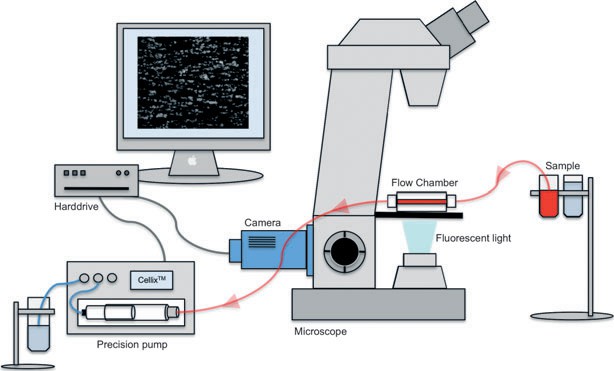 Figure 1. Experimental set-up of the flow chamber-based adhesion assay.[1]
In flow adhesion assays, cells or molecules of interest are labeled with fluorescent dyes or tagged with specific markers to enable their visualization and quantification. The labeled cells or molecules are then introduced into a flow chamber or a microfluidic device, where a controlled flow of fluid is established. The flow rate and shear stress can be adjusted to simulate physiological conditions.
The labeled cells or molecules are perfused over the target surface or substrate, and the adhesion is quantified by monitoring the number of adherent cells or the number of bound molecules. This can be done in real-time using fluorescence microscopy or by capturing images and analyzing them later. Analysis can involve measuring the number of adherent cells, the strength of adhesion, or the distribution of adhesion events.
Flow adhesion assays have broad applications in various fields, including immunology, vascular biology, drug development, and tissue engineering. They are particularly useful in studying cell adhesion molecules, leukocyte adhesion, cancer metastasis, and the interaction of circulating cells with the vascular endothelium. They also serve as valuable tools for evaluating the efficacy of potential therapeutics targeting cell adhesion processes.
At Creative Bioarray, we pride ourselves on providing high-quality Flow Adhesion Assays that are reliable, reproducible, and customizable to meet the unique needs of each research project. With our state-of-the-art microfluidic platforms, we can visualize and quantify cell adhesion events in real-time, providing detailed and quantitative data on cell-cell and cell-substrate interactions.
Study Examples:
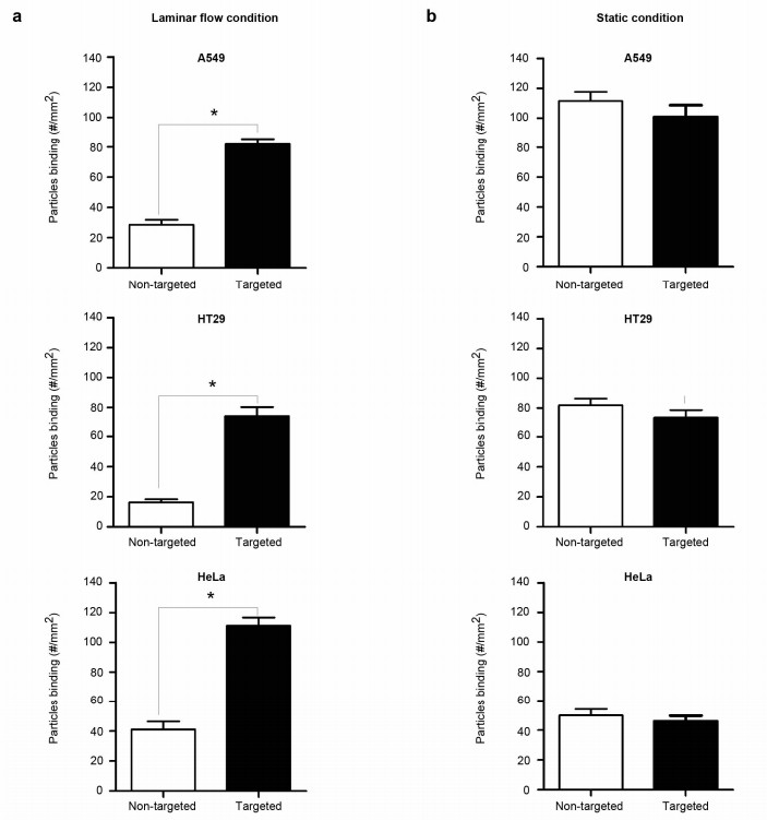 Figure 1. Adhesion of nanoparticles to cancer cells under static or flow conditions. The cell adhesion of nanoparticles in three different cancer cell lines. VEGF-expressing cancer cells were exposed to VEGF-targeted or control nanoparticles either under flow condition (a) or static condition (b). [2]
References:
1. Ferkau A, Ecklebe S, Jahn K, Calmer S, Theilmeier G, Mischke R. A dynamic flow-chamber-based adhesion assay to assess canine platelet-matrix interactions in vitro. Vet Clin Pathol. 2013;42(2):150-156. doi:10.1111/vcp.12035
2. Katawut Namdee, Mattaka Khongkow, Supawadee Boonthod, ect. Cell-based assay for characterizing cell adhesion properties of active targeted nanoparticles under static and flow condition using an integrated flow chamber, Journal of Drug Delivery Science and Technology, Volume 45, 2018, Pages 296-302, ISSN 1773-2247, https://doi.org/10.1016/j.jddst.2018.03.018.
Online Inquiry