
During the chicken embryo development, the chick chorioallantoic membrane (CAM) is formed by fusion of the mesodermal layer of the allantois with the mesodermal layer of the chorion. It is a simple, cost-effective, and efficient model for evaluating the angiogenic potential of various substances such as drugs, growth factors, natural compounds, and more.
To perform the assay, a small window is created in the eggshell above the developing CAM, and a substance of interest is applied directly onto the membrane. The window is then sealed, and the egg is returned to the incubator for a defined period of time. During this time, the substance diffuses into the CAM, and its angiogenic effects can be observed and measured.
Angiogenesis can be evaluated by various methods, including measuring the density and length of blood vessels, counting branching points, assessing vessel density, or analyzing the size of neovascularization areas.
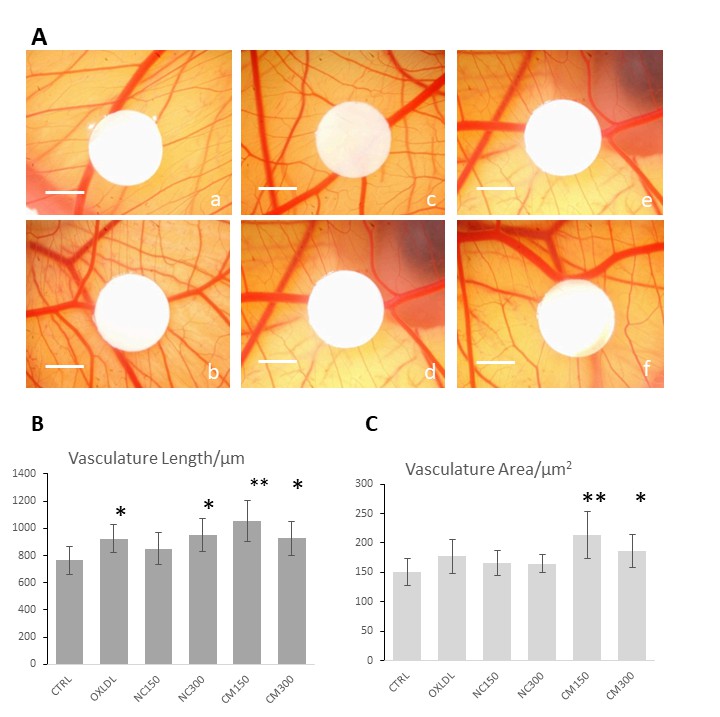 Figure 1. CAM angiogenesis assay showing the pro-angiogenesis effects of the chylomicrons.[1]
Creative Bioarray's CAM Angiogenesis Assay provides researchers with a comprehensive solution for studying angiogenesis. The company offers a wide range of services related to the assay, including customized study design, sample preparation and analysis, and data interpretation.
Reference:
1. WAN, Xiao et al. An Improved In Vivo Angiogenesis Model of Chicken Chorioallantoic Membranes in Surrogate Shells Revealed the Pro-angiogenesis Effects of Chylomicrons. Vascular Cell, [S.l.], v. 11, n. 1, p. 1, mar. 2019. ISSN 2045-824X. doi: http://dx.doi.org/10.24238/13221-11-1-178.
Online Inquiry