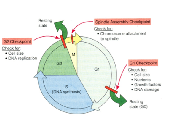
The cell cycle refers to a series of events during cell growth and ends as the cell divides into two daughter cells. Studying the influence of cell cycle changes is important in tumor development and drug development. Creative Bioarray' professional team uses an advanced technology platform to provide a variety of common cell cycle testing services for scientific researchers around the world.
The cell cycle is an important detection parameter. The cell cycle reflects the rate of cell proliferation. From the end of one cell division to the end of the next division is a cycle. The cell cycle usually consists of G0/G1 phase, S phase, G2 phase and M phase.

There are two most important phases in the cell cycle: G1 to S and G2 to M. These two stages are in a period of complex and active molecular level changes, and are easily affected by environmental conditions. If it can be adjusted artificially, it will be of great significance for an in-depth understanding of biological growth and control of tumor growth. The ability to easily and effectively detect cell cycle changes is of great significance to drug development and disease research.
There are many methods to study the cell cycle, including isotope labeling, flow cytometry, and fluorescence detection based on cell imaging. Our services include but are not limited to:
The apoptosis detection provided by Creative Bioarray is based on the "PI and Annexin-V double-label" method. We label Annexin V with fluorescein, use labeled Annexin V as a fluorescent probe, and use a fluorescence microscope or flow cytometer to detect the occurrence of cell apoptosis. PI dye cannot label apoptotic cells and living cells, but it can bind to the DNA inside the cell through the cell membrane of the necrotic cell. Therefore, Annexin V and PI can be used at the same time to distinguish living cells, early apoptotic cells and necrotic cells. Relying on our immunofluorescence imaging platform, Creative Bioarray can provide customers with high-quality image data.

Creative Bioarray provides customers with accurate and reliable flow cytometry PI staining services. The DNA content in the cell is different in each phase of the cell cycle. The fluorescent dye propidium iodide (PI) can bind to DNA in cells. Different amounts of DNA bind to different amounts of fluorescent dye, so the fluorescence intensity detected by flow cytometry is also different, which can reflect the conditions of each phase of the cell cycle.

The DNA content of each phase of the cell cycle is different. Normally, the DNA content of normal cells in the G1/G0 phase is twice that of somatic cells (2N), while the DNA content of cells in the G2/M phase is four times that of somatic cells (4N). The DNA content of S-phase cells is between twice and four times.
This experiment can distinguish the cell cycle phases into G0/G1 phase, S phase and G2/M phase. The cell cycle corresponding to the obtained flow cytometer can be calculated by special software to calculate the cell percentage of each phase.
Fluorescence Detection Service
The FUCCI (fluorescent ubiquitination-based cell-cycle indicator) is a set of fluorescent probes that can visualize cell cycle progression in living cells. The FUCCI technology is based on the identification of two over-expressed proteins geminin and Cdt1 that regulate the cell cycle. Among them, geminin is fused to the green fluorescent group AmCyan, and Cdt1 is fused to the red fluorescent group mCherry. The levels of Cdt1 and geminin constantly fluctuate with changes in the cell cycle: Cdt1 protein levels peak in the G1 phase, while geminin protein levels continue to increase in the S, G2, and M phases. This result is reflected in the FUCCI-expressing cell nucleus in red in the G1 phase and green in the S, G2, and M phases.

By visualizing the cell cycle, FUCCI is a powerful tool for studying any process related to cell growth and differentiation, such as the development and regeneration of organs and cancer.
The time depends on the experiment content
If you are interested in our services, please contact us for more detailed information.
Online Inquiry