
Chemotaxis, the directed movement of cancer cells, inflammatory cells, and stromal cells, is crucial for tumor progression and spread. This process is particularly significant during the metastatic cascade, where cancer cells invade surrounding tissues, enter the bloodstream, exit at a distant location, and establish secondary growth. In healthy cells, chemotaxis is tightly controlled by chemokines, growth factors, and corresponding receptors.[1] However, the chemotactic pathways become dysregulated in cancer progression, enabling metastasis to prevail over normal regulation.
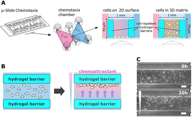 Figure 1. μ-Slide Chemotaxis with a migration arena. A. μ-Slide Chemotaxis was supplemented with a hydrogel migration arena, positioned in the gradient region of the chemotaxis chamber. Dimensions and volumes are indicated or given in the text. B. Function of the migration arena. Cell migration was restricted to the area of the migration arena by barriers from cell-repellent hydrogel. Initially, cells were distributed uniformly in the migration arena. When a gradient was established across the arena, cells migrated in the direction of the chemoattractant and accumulated at one side of the arena. Thus, chemotaxis could be identified from the end-point micrograph. C. Example of keratinocytes distribution in the migration arena at the beginning of the experiment and after 20 hours of migration in a gradient of TGFα. [2]
The μ-Slide Chemotaxis is a specific product developed for conducting chemotaxis assays. These slides are designed to create a controlled environment for studying the migration of cells in response to a chemical gradient. The design typically involves channels or wells that allow the establishment of a gradient of a chemotactic agent across the slide.
The μ-Slide Chemotaxis assay provides high-resolution live-cell imaging capabilities, allowing researchers to track and quantify cell migration in real-time. It is compatible with various imaging modalities, including brightfield, phase-contrast, and fluorescence microscopy.
Creative Bioarray offers a range of μ-Slide Chemotaxis assay services, including different configurations and designs to meet specific research needs. We also provide comprehensive technical support and offer custom services to help researchers design and optimize their experiments.
Study Examples:
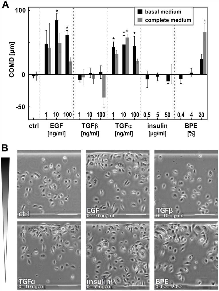 Figure 2. EGF, TGFα and BPE induce directed migration of nHEK. A. nHEK cells were seeded in migration arenas coated with fibronectin. Gradients of EGF, TGFα, TGFβ, insulin, and BPE were established in the chemotaxis chamber, and the effect on cell migration was evaluated after 20 hours.[2]
References:
1. Roussos, E. T., Condeelis, J. S., & Patsialou, A. (2011). Chemotaxis in cancer. Nature Reviews Cancer. 10.1038/NRC3078
2. Tomasova L, Guttenberg Z, Hoffmann B, Merkel R. Advanced 2D/3D cell migration assay for faster evaluation of chemotaxis of slow-moving cells. PLoS One. 2019;14(7):e0219708. Published 2019 Jul 17. doi:10.1371/journal.pone.0219708
Online Inquiry