
The mode of cell migration was originally classified based on the morphology of migration patterns. This terminology was then extended to include molecular parameters, such as cytoskeletal organization. As main categories, cell move either individually (amoeboid or mesenchymal) or collectively (the migration of cohesive multicellular units. The single cell migration is an important part of researches on cell migration and many methods can be used to research it. Among all the methods, Scanning electrochemical microscopy (SECM) is a unique and powerful technique to study single cell migration, which can detect the single-cell movement in three-dimensional (3D) tissue environments.
Scanning electrochemical microscopy (SECM) is an electro-analytical and unique scanning probe technique which can image substrate topography and local reactivity with high resolution. With the ability of quantitative rigor and high sensitivity, the technique has become a popular and wide variety of research applications in the field of biology, corrosion, energy, kinetics and surface modification. Recent advances have greatly increased the capacity of SECM including the ability to characterize interfaces at the nanoscale and to obtain molecular-level chemical information. Thus, the technique of SECM has great advantages in imaging applications compared to other types of scanning probe microscopy. A particular advantage of SECM is that the response observed can be interpreted based on fairly rigorous theory and the measurement can be used to estimate the tip-substrate distance. Moreover, SECM can be employed to image the surfaces of different types of substrates in solution environment.
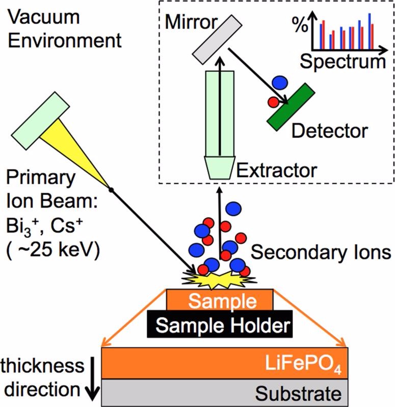 Figure 1: the schematic diagram of Scanning electrochemical microscopy (SECM)
Scanning electrochemical microscopy (SECM) also has great advantages in the study of cell migration, especially in the single cell migration. The newly developed graphite paste ultramicroelectrode (UME) showed significantly less fouling and background, thus it could be used to monitor and track the migration pattern of a single cell. The technique of SECM can also be employed to measure the morphology of a single live cancer cell during cellular migration and determined these dimensions by the probe scan curves. Moreover, this non-invasive SECM-based technique could potentially be expanded to other cell lines to obtain an improved understanding of the structure–function relationship at the level of a single cell by studying cellular biomechanics.
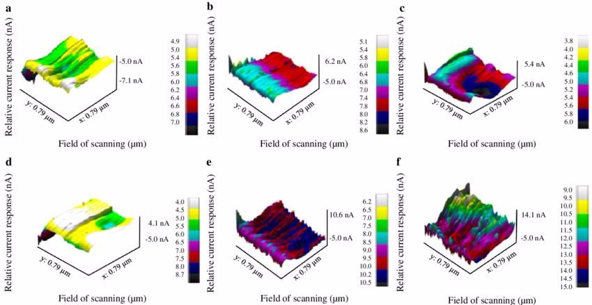 Figure 2: The experimental results of Scanning Electrochemical Microscopy (SECM)

*If your organization requires signing of a confidentiality agreement, please contact us by email.
Online Inquiry