
As proliferating neoplastic cells attempt to escape the primary tumor site, local cell adhesion and invasion of the surrounding tissue must occur [1]. Prior to penetrating the blood vessel endothelium and gaining access to the blood stream, cancer cells must invade local tissues by degrading ECM protein components and ultimately, transverse the basement membrane. Once in circulation, these cells can form metastatic colonies at secondary locations [2].
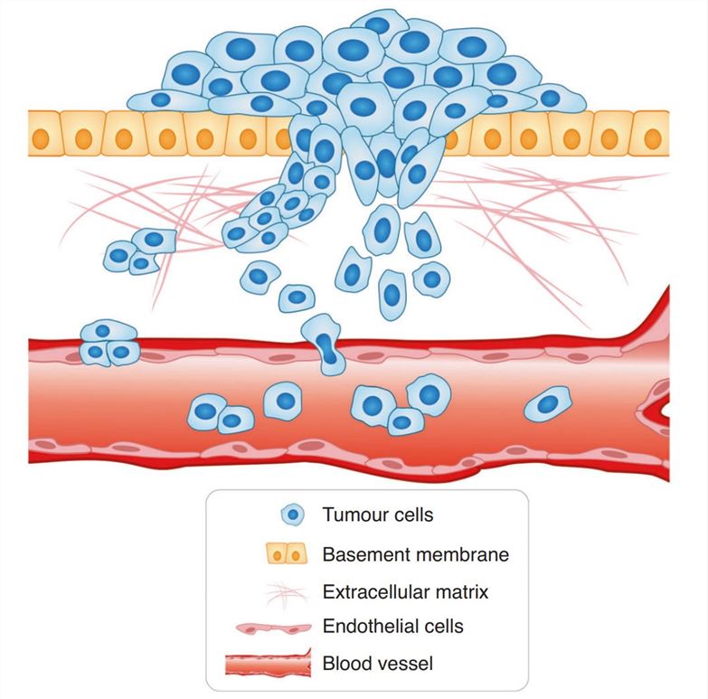 Figure 1. The model of cancer cell invasion.
Cell invasion assays have been used to study the interactions between tumor cells and the extracellular matrix (ECM), which not only provides a structural scaffold for the cell, but also contains various biological factors for the survival and growth of the cell. ECM gel contains the basic components of the ECM that provides a structural support for the cell to grow and move. Cells can secrete enzymes that degrade certain components of the ECM to move towards chemoattractants, or to simply establish niches for growth. Metastatic tumor cells often show more invasiveness to the ECM gel due to their higher motility and/or enzymatic activity for degrading ECM components.
Creative Biarray provides a range of services related to cell invasion assays, including the Transwell Invasion Assay and 3D Spheroid Invasion Assay.
References:
1. Friedl, Peter, and Stephanie Alexander. "Cancer invasion and the microenvironment: plasticity and reciprocity." Cell vol. 147,5 (2011): 992-1009. doi:10.1016/j.cell.2011.11.016
2. Novikov, Nikita M et al. "Mutational drivers of cancer cell migration and invasion." British journal of cancer vol. 124,1 (2021): 102-114. doi:10.1038/s41416-020-01149-0
Online Inquiry