The quantification of cell migration rate is an important part in the research of the modulatory potential of extrinsic factors on cancer cell migration and quick resurfacing. It is essential for intestinal wound repair to regain the ability to function as a protective barrier. Different techniques have been employed to quantify cell migration. One of the most widely used techniques is cell exclusion zone assay which can get precise estimates of cell migration and allow continuous visual assessment of the cells by the ability to acquire multiplexed data without the observation restrictions.
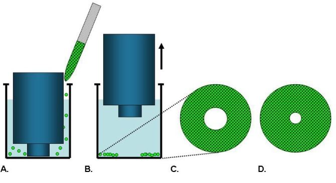
 Figure 1. The basic experimental process of cell exclusion zone assay
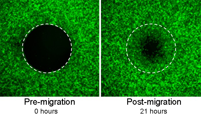
Cell exclusion zone assay is especially appropriate for studying cell migration on an uncompromised surface uncoupled from contributions of cell damage and permeabilization. To avoid damaging cells and matrix, cell-free areas are created in cell exclusion zone assay by that cells are seeded around removable silicone stoppers which exclude cells from attaching to a central zone. The cell density is adjusted to form a fully confluent. After cell adhesion, the stoppers are removed to reveal a circular cell-free area, wherein the cells will migrate. Then, upon the appropriate staining with fluorescent dyes and microscopically visualizing the monolayers, the migration rate is quantified by counting the cells which have migrated to the circular cell-free area.
 Figure 2. The experimental results of cell exclusion zone assay
Cell exclusion zone assay has many distinct advantages in the research of cell migration, which can acquire continuous visual assessment and multiplexed data without the restrictions of observation. In this way, information regarding morphology, distance, velocity and direction of migration can be collected. In addition, cell exclusion assay offers robust and reproducible data because the detection zone are accurately and precisely positioned in the assay.
There are several sophisticated equipment in our advanced platform including high throughput imaging system which can continuously observe living cells. Several attempts have been made to optimize the assay by our specialists. Thus, we can offer higher throughput and accurate experimental results automated and advanced imaging system, which can offer reproducible results with consistence.

*If your organization requires signing of a confidentiality agreement, please contact us by email.
Online Inquiry