Pyroptosis, a caspase-dependent inflammatory cell death, is marked by pore formation, cell swelling, plasma membrane rupture, and the release of intracellular contents.

Pyroptosis: A new frontier in cancer. 
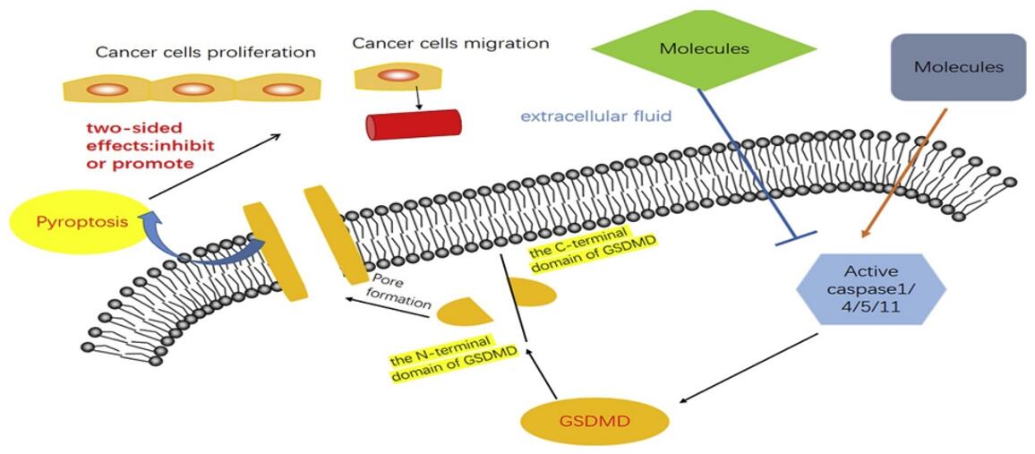
Figure 1. Pyroptosis: A new frontier in cancer. [1]

Pyroptosis is distinguished by the N-terminal cleavage of Gasdermin D (GSDMD), leading to its oligomerization and the formation of a lytic pore in the plasma membrane. This cleavage event is mediated by inflammatory caspases 1, 4, 5, and 11, distinct from those involved in apoptosis. Monitoring the cleavage of GSDMD and its related family members, along with these specific inflammatory caspases, is essential for a comprehensive study of pyroptosis. Recent studies have revealed that pyroptosis, influenced by non-coding RNAs and various molecules, plays a pivotal role in regulating the proliferation, invasion, and metastasis of tumors.

Creative Bioarray offers an advanced pyroptosis assay, a state-of-the-art solution for exploring and understanding the intricate mechanisms of pyroptosis in cancer cells. Our assay provides a robust platform to investigate pyroptosis induction and its impact on various cancer cell types.

Methods for Monitoring Cancer Cell Pyroptosis

  • Gasdermin D Cleavage Assays
    Western Blotting: Detect cleaved fragments of gasdermin D (GSDMD) using specific antibodies to assess pyroptosis induction.
    Immunoblot Analysis: Monitor the appearance of N-terminal fragments of GSDMD, indicative of its activation during pyroptosis.
  • Pore Formation Assays
    Propidium Iodide Uptake: Measure the influx of propidium iodide into cells, indicating the formation of pores in the plasma membrane.
    Fluorescent Dyes: Use membrane-impermeable dyes, such as YoPro-1 or ethidium homodimer-1, to assess pore formation.
  • Release of Pro-Inflammatory Cytokines
    ELISA: Quantify the release of pro-inflammatory cytokines (IL-1β, IL-18) into the culture medium as an indicator of pyroptosis.
  • Cell Morphology and Imaging
    Microscopy: Observe cellular morphological changes, including cell swelling and membrane rupture, using phase-contrast or fluorescence microscopy.
    Transmission Electron Microscopy (TEM): Visualize ultrastructural changes associated with pyroptosis, such as membrane disruption and cytoplasmic swelling.
  • Caspase Activation Assays
    Immunofluorescence Assays: Measure the activity of specific inflammatory caspases (e.g., caspase-1) using fluorogenic substrates.
    Western Blotting: Detect cleaved caspase fragments as an indicator of caspase activation during pyroptosis.
  • Cell Viability and Cytotoxicity Assays
    MTT Assay or Alamar Blue Assay: Measure changes in cell viability as an indirect indicator of pyroptosis-induced cytotoxicity.

References:

1. Fang Y, Tian S, Pan Y, et al. Pyroptosis: A new frontier in cancer. Biomed Pharmacother. 2020;121:109595. doi:10.1016/j.biopha.2019.109595

2. Wang S, Liu Y, Zhang L, Sun Z. Methods for monitoring cancer cell pyroptosis [published online ahead of print, 2021 Dec 22]. Cancer Biol Med. 2021;19(4):398-414. doi:10.20892/j.issn.2095-3941.2021.0504

* For scientific research only

Online Inquiry