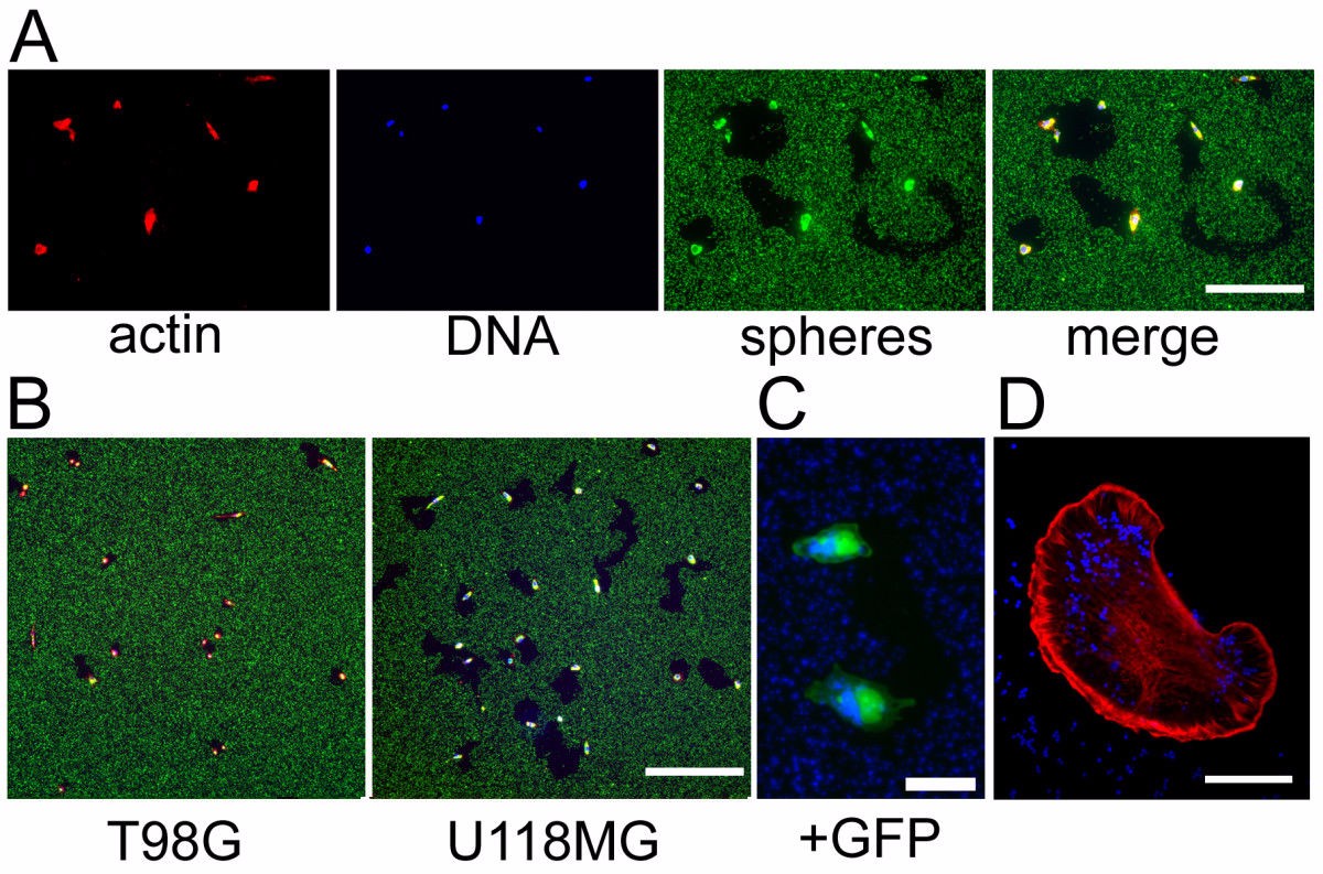
Identifying and characterizing cell behaviors will facilitate the understanding of the molecular basis regulating these behaviors. Although many approaches exist to characterize cell mobility, there are still many changes in analyzing cell mobility, especially for the specific cell mobility. A novel and versatile method is described below to study cell mobility including the specific cell mobility.
In this approach, we take advantage of the phagokinetic ability of migrating cells to label the migrating cells with fluorescent dyes. First, non-cytotoxic fluorescent polystyrene microspheres are utilized as cell labels and coated onto a variety of migratory substrates in tissue culture vessels. Cells are seeded onto the pre-treated tissue culture plastic. Then cells will be fixed with 4% paraformaldehyde after incubated for appropriate time. Moving cells will generate microsphere-free areas in the dense fluorescent particle coat that are easily visualized using fluorescence microscopy, since the migrating cells can phagocytize the fluorescent microspheres during the process of cell mobility. The paths of cell migration can be easily tracked and we can quantify cell migration both by direct measurement of cleared area and by fluorescent signal intensity within single cells, and permits isolation of cells based on their mobility. High quality data can be obtained in this assay by evaluating cleared area and fluorescent signal simultaneously. The real-time path of cell migration can be monitored and the absolute speed of cell migration can be determined by high throughput living image system.

Since this method is based on the fluorescent label to study cell migration, it also called fluorescent-labeled cell migration assay. This method enables recovery of characterized cell populations, and can enable screens to identify factors that might regulate mobility differences even with clonal population of cells. It also creates value insights to understand the molecular mechanisms of cell migration. In addition, we can stain migrating cells with distinct fluorescent markets and utilize beads of different fluorescent emission wavelength to track cell migration simultaneously. Thus, we can compare the mobility of different types of cell lines simultaneously, it is of great advantage to study the characteristic of cell mobility of different cell lines and study the potential molecular pathways that regulate the behavior of interest. This assay is also highly sensitive-single cell characteristics, including the behavior of individual transfected cells, can be obtained in an unbiased fashion.

*If your organization requires signing of a confidentiality agreement, please contact us by email.
Online Inquiry