
Blood vessels deliver blood to all organs and tissues of the body, supplying oxygen and nutrients. When the local demands could no longer be met, cells secrete vascular endothelial growth factor (VEGF) to stimulate pre-existing vessels to form new blood vessels. This process is known as angiogenesis, which plays an essential role in organ and tissue development, regeneration, and tumor progression. When the angiogenesis process is dysregulated, a number of pathological conditions can arise. Therefore, drugs affecting angiogenesis show great promise in mitigating angiogenesis-dependent diseases, such as cancer, diabetic retinopathy, and neovascular age-related macular degeneration (NVAMD).
A three-dimensional (3D) spheroid angiogenesis assay system has been developed to mimic angiogenic responses and sprouting behavior, which can be used to assess the effects of drugs on angiogenesis. Creative Bioarray offers 3D angiogenesis assay to assist you in investigating angiogenesis responses to test compounds and identify potential drug candidates.
To produce spheroids, endothelial cells (ECs) are cultured as hanging drops. The spheroids are then embedded in collagen gels. EC spheroids can form capillary-like sprouts which are relatively close to the physiological angiogenesis. By analyzing the number and length of sprouts, the effect of drug treatment on angiogenesis can be quantified.
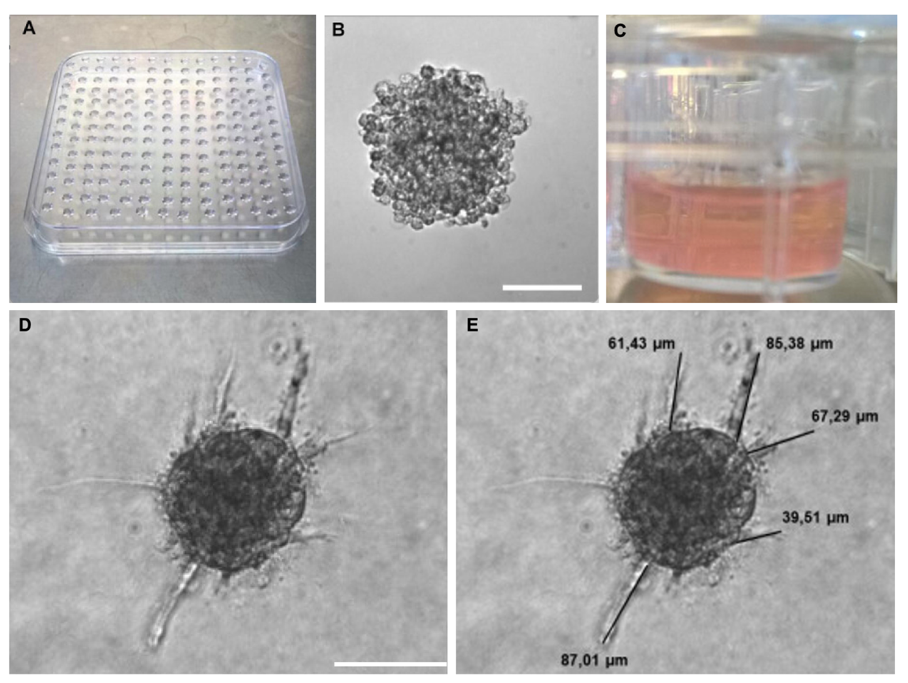 Figure 1. The procedure of 3D angiogenesis assay. A) Petri dish with hanging drops to form spheroids. B) Representative image of a spheroid in a hanging drop after 24 h of upside-down incubation. Scale bar, 100 µm. C) Spheroids are embedded in collagen gels. D) Image of a human umbilical vein endothelial cell (HUVEC) spheroid embedded in collagen gels after 24h of sprouting. Scale bar, 100 µm. E) Image analysis. The sprout length of all sprouts in a spheroid is measured by FIJI software.
A major advantage of this technique is the possibility to analyze angiogenesis in a 3D system, which resembles more closely to in vivo conditions. This promotes cell-cell signaling between endothelial cells. Moreover, this assay mimics the entire process of angiogenesis in vitro, including proteolytic degradation, cell migration, proliferation, and lumen formation, all in three dimensions. Therefore, spheroid-based angiogenesis assay is an effective in vitro assay for evaluating pro- or anti-angiogenic properties.
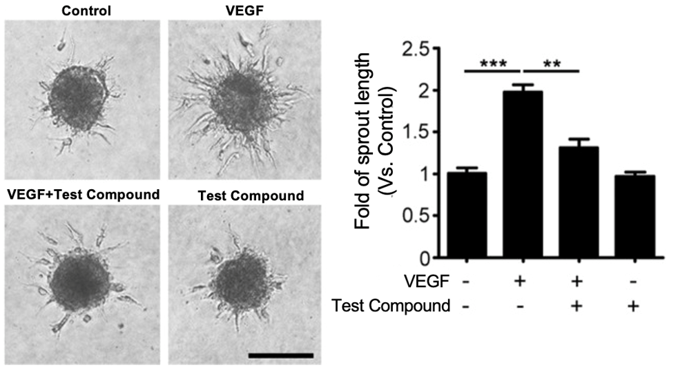 Figure 2. The test compound suppressed VEGF-induced spheroid sprouting of HUVECs. Scale bar, 200 µm.
References:
1. Tetzlaff, Fabian, and Andreas Fischer. "Human endothelial cell spheroid-based sprouting angiogenesis assay in collagen." Bio-protocol 8.17 (2018): e2995-e2995.
2. Heiss, Maximilian, et al. "Endothelial cell spheroids as a versatile tool to study angiogenesis in vitro." The FASEB Journal 29.7 (2015): 3076-3084.
3. Seo, Songyi, et al. "Evogliptin, a dipeptidyl peptidase-4 inhibitor, attenuates pathological retinal angiogenesis by suppressing vascular endothelial growth factor-induced Arf6 activation." Experimental & molecular medicine 52.10 (2020): 1744-1753.
Online Inquiry