Creative Bioarray is dedicated to providing customers with a professional cell biology research platform. We have an experienced team of experts who can provide you with the most professional services in the research of cell death, saving your time and efforts.
Mitochondria play a pivotal role in the process of apoptosis. Changes in mitochondrial membrane potential and membrane permeability are important features of apoptotic cells.
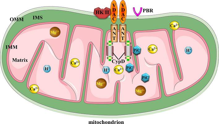
In the process of cell death, the mitochondrial permeability transition pore (MPTor MPTP) significantly changes the permeability of mitochondria. After MPTP is activated, the mitochondrial membrane potential decreases and cytochrome C is released.
 Figure1. The structure of mitochondrial membrane permeability transition pore (Li Y, et al, 2020)
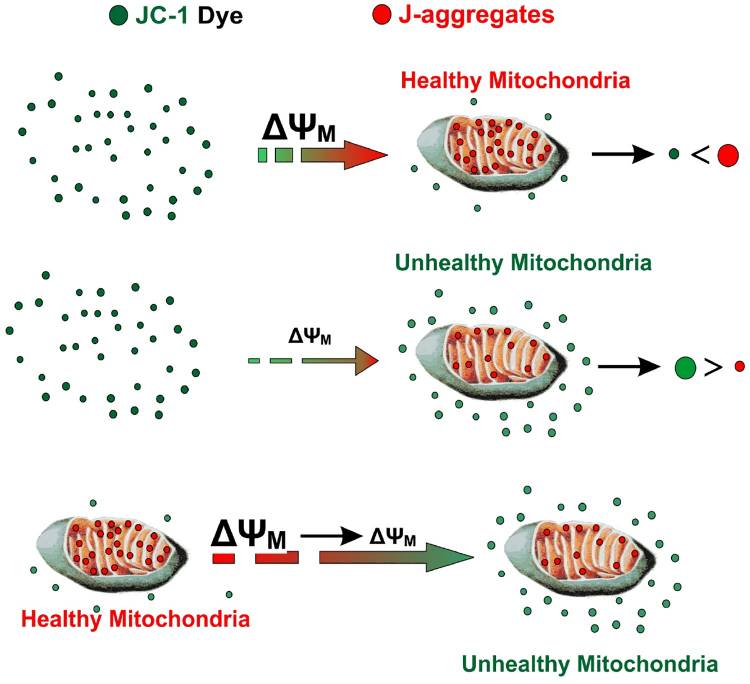
The decrease of mitochondrial transmembrane potential is considered to be the earliest in the process of apoptosis cascade. JC-1 is an ideal fluorescent probe that is widely used to detect mitochondrial membrane potential.
 Figure2. JC-1 enters the mitochondria and produces J aggregates (Sivandzade F,et al, 2019)
We provide a variety of cell death-related mitochondrial analysis services to meet your needs. Our services include but are not limited to:
Creative Bioarray provides you with high-quality cytochrome C and membrane permeability test kits and different detection methods such as fluorescence microscope and flow cytometry testing services to help your cell death research such as the studies of apoptosis and necrosis.
MPTP is a non-specific channel composed of the inner and outer mitochondrial membranes. It is believed to be involved in the release of cytochrome C in cell death. There are high-quality cytochrome C and membrane permeability test kits and corresponding testing services available for your choice.
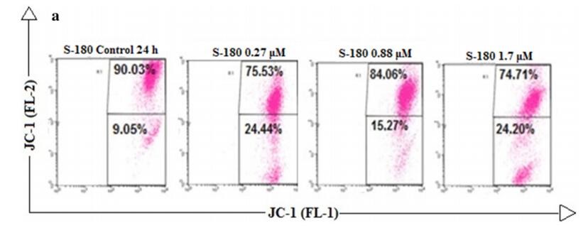
Creative Bioarray uses the JC-1 fluorescent probe method to detect the mitochondrial membrane potential. The change of the fluorescence signal of the JC-1 probe to detect the change of the mitochondrial membrane potential in apoptotic cells is a quick, simple and effective detection method, which can be used for flow cytometric analysis, fluorescence microscope observation and 96-well fluorescence microplate reading.
 Figure3.Use JC-1 dye to detect mitochondrial membrane potential by flow cytometry(Wanessa Carvalho Pires, et al, 2017)
When the mitochondrial membrane potential is high, JC-1 aggregates in the matrix of the mitochondria to form a polymer, which can produce red fluorescence; when the mitochondrial membrane potential is low, JC-1 cannot accumulate in the matrix of the mitochondria to produce green fluorescence.
The time depends on the experiment content
If you are interested in our services, please contact us for more detailed information.
References:
Online Inquiry