
The process of remyelination can be divided into two primary stages. Initially, oligodendrocyte progenitor cells (OPCs) are recruited through proliferation and potentially migration. Subsequently, these OPCs interact with demyelinated axons and transform into oligodendrocytes that form myelin sheaths. It is possible for the remyelination process to fail at either of these stages, both of which become less effective with aging, which likely contributes to the decline in remyelination efficiency observed during the disease.
Creative Bioarray's in vitro model of proliferating OPCs is a valuable tool for assessing how specific stimuli directly affect OPC proliferation and survival.
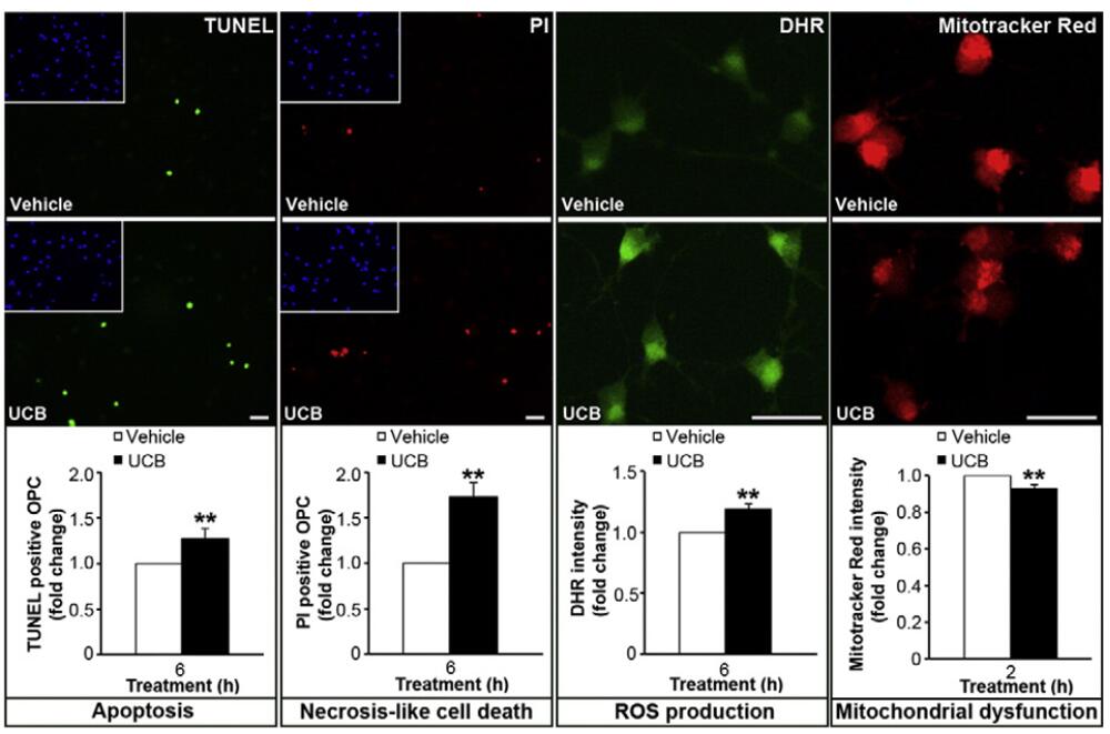 Figure 1. Model of Oligodendrocyte Proliferation and Potential Evaluations.
In this model it is possible to incubate OPC with different compounds, like unconjugated bilirubin (UCB), to evaluate and compare changes in OPC viability
by TUNEL assay or propidium iodide (PI) or mechanisms underlying OPC demise. By fluorescence it is possible to evaluate individual cell production of
reactive oxygen species (ROS) using dihydrorhodamine 123 (DHR) dye or mitochondrial dysfunction using Mitotracker® Red dye.[1]
Figure 1. Model of Oligodendrocyte Proliferation and Potential Evaluations.
In this model it is possible to incubate OPC with different compounds, like unconjugated bilirubin (UCB), to evaluate and compare changes in OPC viability
by TUNEL assay or propidium iodide (PI) or mechanisms underlying OPC demise. By fluorescence it is possible to evaluate individual cell production of
reactive oxygen species (ROS) using dihydrorhodamine 123 (DHR) dye or mitochondrial dysfunction using Mitotracker® Red dye.[1]
Reference:
1. Barateiro A, Fernandes A. Temporal oligodendrocyte lineage progression: in vitro models of proliferation, differentiation and myelination. Biochim Biophys Acta. 2014;1843(9):1917-1929. doi:10.1016/j.bbamcr.2014.04.018.
Online Inquiry