Creative Bioarray uses the morphological changes of nuclear chromatin as an indicator to determine the stages of apoptosis. The identification of classic apoptosis indicators is usually based on morphology.

When apoptosis occurs, the cell shrinks, the cytoplasm is dense, and the nuclear chromatin is bordered. The nucleus is then lysed, and the cytoplasmic buds fall off, forming membrane-coated apoptotic bodies containing nuclear debris and/or organelle components. Hoechst and DAPI are popular blue fluorescent, nuclear-specific dyes that can be used to stain live or fixed cells. The staining is very stable and non-toxic to live cells for several days or longer.
We provide a variety of apoptosis morphology detection services to meet your needs. Our analytical methods include but are not limited to:
Electron Microscopy
Creative Bioarray uses transmission electron microscopy to observe cell structure changes in different periods of apoptosis. Our professional scientists use advanced equipment to provide high-resolution images and analysis reports.
Apoptosis at different stages has different and prominent characteristics. Morphological observation by electron microscopy is by far the most classic and reliable method for studying apoptosis. In articles focusing on apoptosis, electron microscopy data is necessary, especially for mainstream journals.
DNA Specific Staining Service
In addition to the use of electron microscopy, DNA-specific staining has also become one of the most commonly used detection indicators for apoptosis. However, it often takes a lot of time and labor costs to quantify the image information. Creative Bioarray provides a one-stop service from sample staining to quantifying results, saving your time and cost. The common staining we use are Hoechst and DAPI.
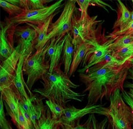
The fluorescent Hoechst stain can enter the normal cell membrane in a small amount, making it light blue. The membrane permeability of apoptotic cells is enhanced, and the structure of chromosomal DNA changes, so more Hoechst can enter the apoptotic cells with enhanced blue fluorescence.
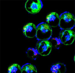
DAPI can penetrate intact cell membranes to stain live and fixed cells. Because of its high affinity for DNA, it is frequently used for counting cells, measuring apoptosis, sorting cells based on DNA content, and as a nuclear segmentation tool in high-content imaging analysis.
The time depends on the experiment content
If you are interested in our services, please contact us for more detailed information.
Online Inquiry