Neuroinflammatory refers to inflammation that occurs in the brain or spinal cord. This inflammation can be caused by a variety of factors, including infections, traumatic injuries, autoimmune disorders, and chronic neurodegenerative diseases.
Neuroinflammation involves the activation of various immune cells within the central nervous system (CNS), including microglia and astrocytes. Additionally, several signaling molecules, such as cytokines and chemokines, play crucial roles in mediating and regulating the inflammatory response.
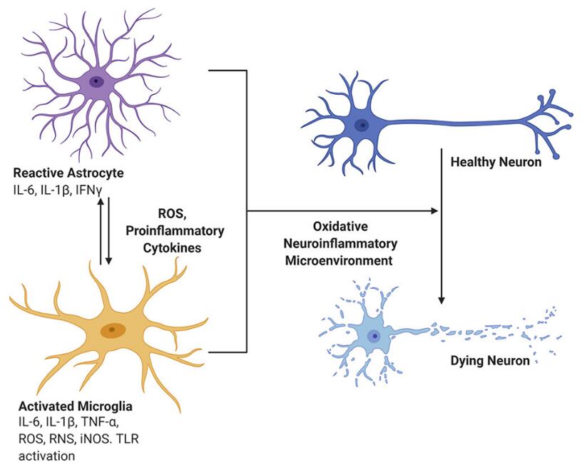 Figure 1. Role of proinflammatory mediators in neurodegeneration: Production of inflammatory mediators such as cytokines and chemokines is central for the development of the neuroinflammatory microenvironment in neurodegenerative diseases, demyelinating diseases, and other neurological disorders. [1]
Lipopolysaccharide (LPS) is a component of the cell wall of Gram-negative bacteria, and it is commonly used to induce an inflammatory response in cell cultures, including those of neuronal cells. Creative Bioarray offers LPS-induced neuroinflammation cellular model to assess the efficacy of potential anti-neuroinflammation drugs. Our experienced team of scientists can design and conduct experiments using various cell lines or primary cells to study the effects of test compounds on inflammatory markers, cytokine release, cell viability, and other relevant endpoints.
Study Examples:
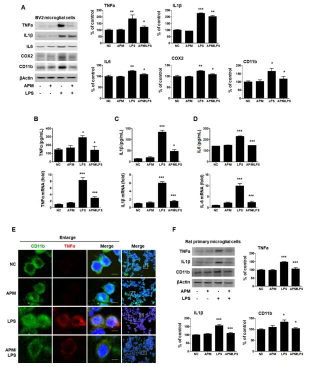 Figure 2. APM inhibits LPS-induced proinflammatory responses in LPS-stimulated BV2 and rat primary microglial cells. Cells were incubated in the presence or absence of APM for 1 h and then treated with LPS for 12 h. [2]
References:
1. Kaur, Navrinder et al. "Neuroinflammation Mechanisms and Phytotherapeutic Intervention: A Systematic Review." ACS chemical neuroscience vol. 11,22 (2020): 3707-3731. doi:10.1021/acschemneuro.0c00427
2. Park, Jihyun et al. "Apamin Suppresses LPS-Induced Neuroinflammatory Responses by Regulating SK Channels and TLR4-Mediated Signaling Pathways." International journal of molecular sciences vol. 21,12 4319. 17 Jun. 2020, doi:10.3390/ijms21124319
Online Inquiry