Drug-induced cardiotoxicity assays are essential tools in preclinical drug development to assess the potential adverse effects of pharmaceutical compounds on the cardiovascular system. These assays aim to identify any detrimental impact on the heart, including effects on cardiac cells, tissues, and function.
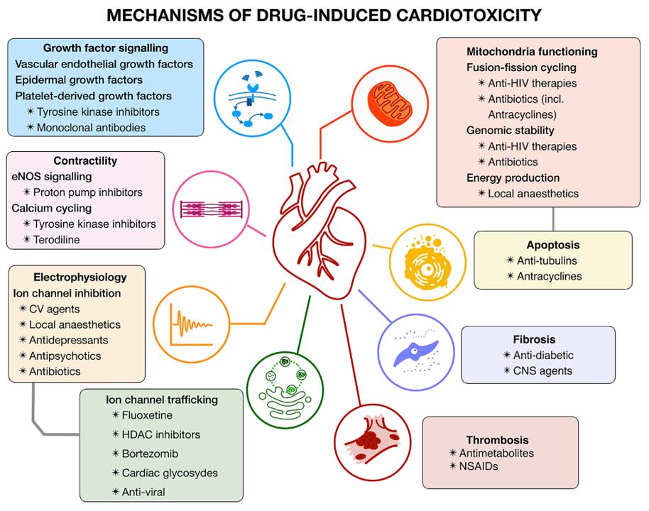 Figure 1. Mechanisms of drug-induced cardiotoxicity [1]
In vitro Cellular Models
- 2D models: iPSC- / ES-derived cardiomyocytes
- 3D cardiotoxicity assay: With 3D cardiomyocyte spheroids and high-content imaging devices, Creative Bioarray can provide a cardiotoxicity service that monitors the effects of compounds on cardiomyocytes at a structural and phenotypic level.
In vitro drug-induced cardiotoxicity assays
- Cell Viability and Cytotoxicity Assays:
- MTT Assay: Measures mitochondrial activity as an indicator of cell viability.
- LDH Release Assay: Detects the release of lactate dehydrogenase, indicating cytotoxicity and cell membrane damage.
- Electrophysiological Assays:
- Patch clamp assays: Patch clamp assays are used to measure the activity of ion channels in cardiac cells. Changes in ion channel activity can indicate potential cardiotoxic effects of a drug.
- Mitochondrial Function Assays:
- Mitochondrial Membrane Potential (MMP) Assay: Assesses drug-induced changes in mitochondrial function.
- ATP Assay: Measures cellular ATP levels, reflecting overall cellular energy status.
- Oxidative Stress Assays:
- Reactive Oxygen Species (ROS) Assay: Measures the production of reactive oxygen species, indicating oxidative stress.
- Glutathione Assay (GSH): Assesses changes in cellular antioxidant capacity.
Overall, in vitro drug-induced cardiotoxicity assays are valuable tools for assessing the safety of new pharmacological compounds and can help to identify potential cardiotoxic effects early in the drug development process.
Reference:
1. Mamoshina, Polina et al. "Toward a broader view of mechanisms of drug cardiotoxicity." Cell reports. Medicine vol. 2,3 100216. 16 Mar. 2021, doi:10.1016/j.xcrm.2021.100216
* For scientific research only